Shadow Imaging
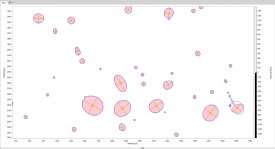
High-magnification shadow Imaging is very suitable for visualizing particles, droplets and other structures. The technique is based on high resolution imaging with pulsed backlight illumination. The measurement volume is defined by the focal plane and the depth of field of the imaging system. This technique is independent of the shape and material (either transparent or opaque) of the particles and allows the investigation of sizes down to the micro scale using an appropriate optical system.

The light source can be a pulsed laser with special illumination optics or a flash lamp, depending on the velocity of the particles. Using a short laser pulse as illumination, it is possible to "freeze" motions of more than 100 m/s. A double pulse laser combined with a double frame camera allows the investigation of size dependent particle velocities.

Interferometric Mie Imaging: IMI
(aka Interferometric Laser Imaging for Droplet Sizing, ILIDS)
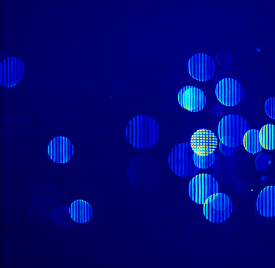
Interferometric Mie Imaging (IMI) is a sizing technique for the evaluation of the diameter of spherical particles, droplets and bubbles similar to PDI. The working principle is based on the out-of-focus imaging of particles illuminated by a laser light sheet. The optical setup of a standard IMI system consists of a laser-light sheet and a digital camera with a high quality lens. Moving the camera chip to an out-of-focus position an interference fringe pattern becomes visible. The visible fringe pattern corresponds exactly to the far field scattering which can be calculated by the Mie theory. The number of fringes within the aperture image depends on the diameter of the droplet and the aperture angle. With increasing particle size, the number of fringes increases. The exact size of the particles is determined by analysis of the fringe patterns. The size of the aperture image is a measure for the z-position of the particle along the line-of-sight.

